BS-2094C Inverted Biological Microscope
Introduction
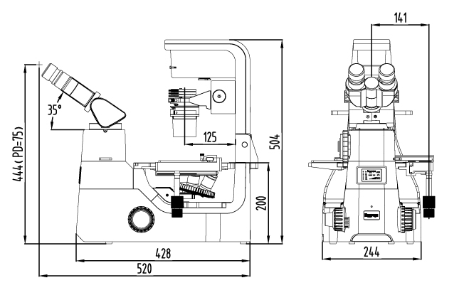
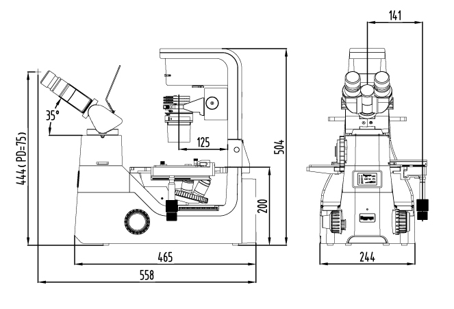
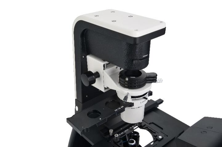
BS-2094C Inverted Biological Microscope is a high level microscope which is specially designed for medical and health units, universities, research institutes to observe cultured living cells. With innovative infinite optical system and ergonomic design, it has excellent optical performance and easy to operate features. The microscope has adopted long life LED lamps as transmitted and fluorescent light source. Digital cameras can be added to the microscope on left side to take photos, videos and make measurement. The tilting head can offer a comfortable working mode. The angle of transmitted illumination arm can be adjusted, so petri-dish or flask can be easily moved out.
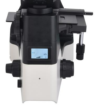
BS-2094C has an intelligent illumination management system, the illumination intensity will automatically change after you change the objectives and make the microscope to get the best illumination effect, BS-2094C also has a LCD screen to show the working mode like magnification, light intensity, transmitted or fluorescent light source, working or sleep etc.
Feature
2. Camera port is on left side, less disturb for operation. Light distribution (both): 100 : 0 (100% for eyepiece); 0 : 100 (100% for camera).
3. Long working distance condenser N.A. 0.30, Working distance: 75mm(with condenser).
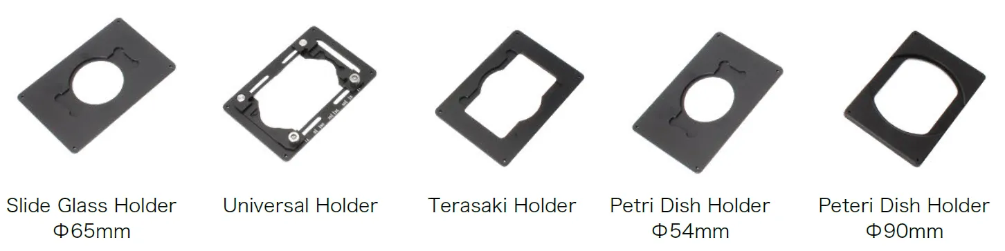
4. Large size stage, convenient for research. Stage Size: 170mm(X) × 250 (Y)mm, Mechanical stage moving range: 128mm (X) × 80 (Y)mm. Various of petri-dish holders are available.
(1) Coded Quintuple Nosepiece can memorize the illumination brightness of each objective. When different objectives are converted to each other, the light intensity is automatically adjusted to reduce visual fatigue and improve work efficiency.
Double click: light intensity lock or unlock
Rotation: Adjust brightness
Press + clockwise rotate: Switch to the transmitted light source
Press + contrarotate: Switch to the fluorescent light source
Press 3 seconds: Set the time of turning off the light after leaving
The LCD screen in the front of the microscope can display the working mode of the microscope, including magnification, light intensity, sleep mode and so on.

(1) Phase contrast observation is a microscopic observation technique that produces a high-contrast microscopic image of a transparent sample by utilizing a change in refractive index. The advantage is that the details of live cell imaging can be obtained without staining and fluorescent dyes.
Application range: Living cells culture, Micro-organism, Tissue slide, cell nuclei and organelles etc.
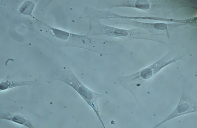
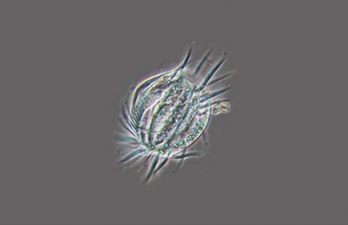
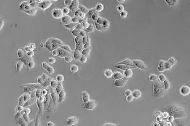
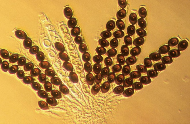
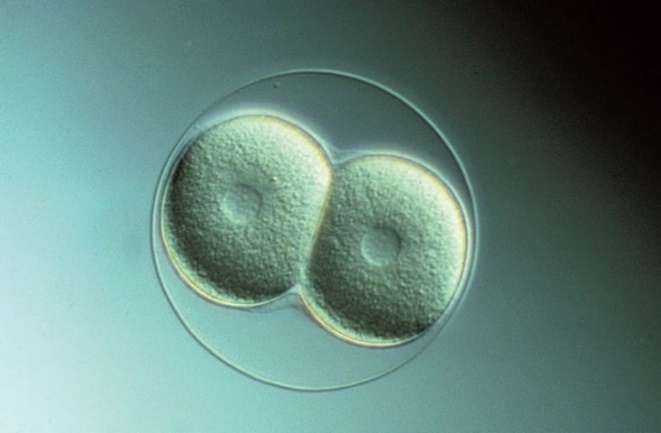
With Hoffman Modulation Phase Contrast With 3D Emboss Contrast
9. LED Fluorescent attachment is optional.
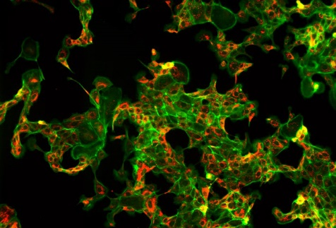
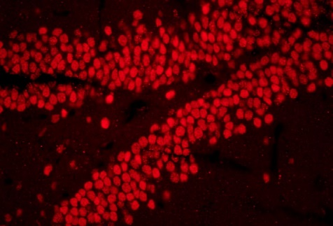
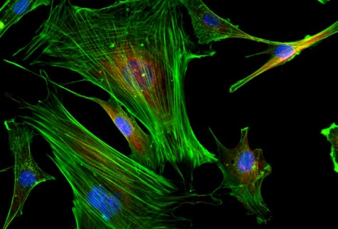
(1) LED light makes fluorescent observation easier.
Fly-eye lens and Kohler illumination have provided a uniform and bright field of view, which is benefit to get high definition images and perfect details. Compared with traditional mercury bulb, the LED lamp has much longer working life, it saves money and has greatly improved the working efficiency. The problems of preheating, cooling and high temperature of mercury lamp has also been solved.
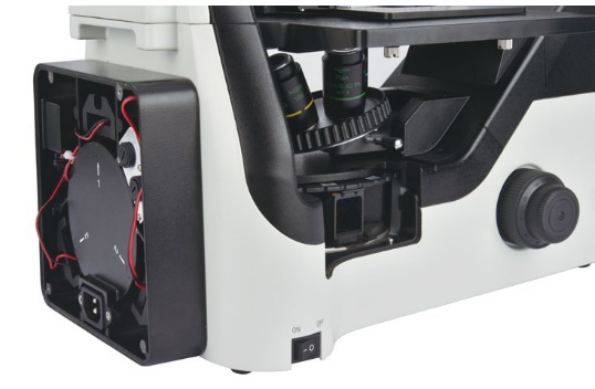
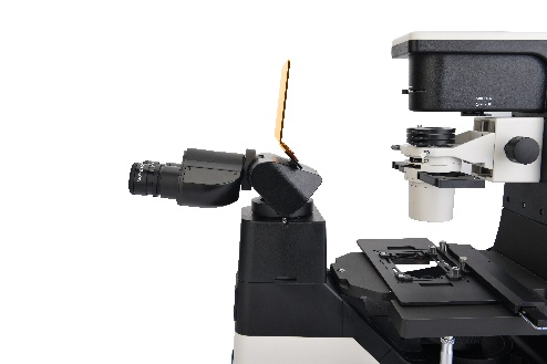
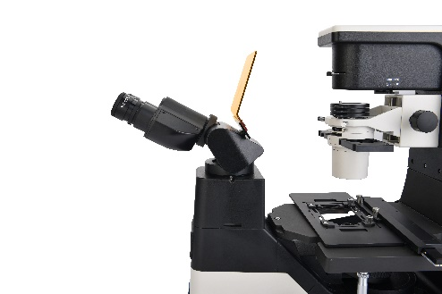
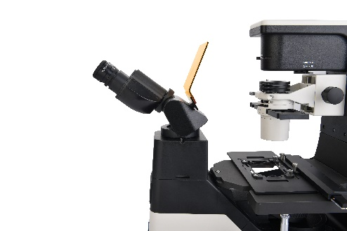
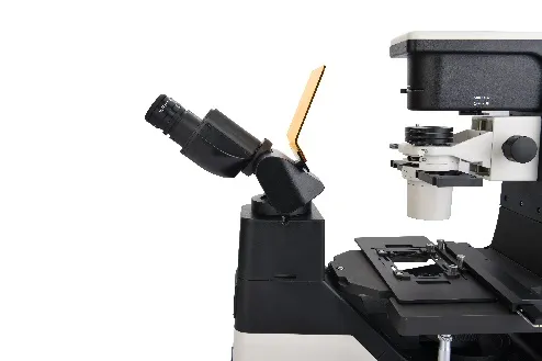
The culture dishes used for cell observation often have a larger volume and area, and the tiltable transmitted illumination column provides more space for sample replacement, which is more convenient for users to operate.
Application
BS-2094C inverted microscope can be used by medical and health units, universities, research institutes for observations of micro-organisms, cells, bacteria and tissue cultivation. They can be used for continuous observation of process of cells, bacteria grow and divide in the culture medium. Videos and images can be taken during the process. These microscopes are widely used in cytology, parasitology, oncology, immunology, genetic engineering, industrial microbiology, botany and other fields.
Specification
Item | Specification | BS-2094C | BS-2094CF | |
Optical System | NIS 60 Infinite Optical System, Tube length 200mm | ● | ● | |
Viewing Head | Seidentopf Tilting Binocular Head, adjustable 5-35° inclined, Interpupillary Distance 48-75mm, Left side camera port, Light distribution: 100: 0 (100% for eyepiece), 0:100 (100% for camera), Eyepiece Tube Diameter 30mm | ● | ● | |
Eyepiece | SW10×/ 22mm | ● | ● | |
WF15×/ 16mm | ○ | ○ | ||
WF20×/ 12mm | ○ | ○ | ||
Objective (Parfocal distance 60mm, M25×0.75) | NIS60 Infinite LWD Plan Achromatic Objective | 4×/0.1, WD=30mm | ● | ○ |
10×/0.25, WD=10.2mm | ○ | ○ | ||
20×/0.40, WD=12mm | ○ | ○ | ||
40×/0.60, WD=2.2mm | ○ | ○ | ||
NIS60 Infinite LWD Plan Phase Contrast Achromatic Objective | PH10×/0.25, WD=10.2mm | ● | ○ | |
PH20×/0.40, WD=12mm | ● | ○ | ||
PH40×/0.60, WD=2.2mm | ● | ○ | ||
NIS60 Infinite LWD Plan Semi-APO Fluorescent Objective | 4×/0.13, WD=17mm, cover glass=- | ○ | ● | |
10×/0.3, WD=7.4mm, cover glass=1.2mm | ○ | ● | ||
20×/0.45, WD=8mm, cover glass=1.2mm | ○ | ● | ||
40×/0.60, WD=3.3mm, cover glass=1.2mm | ○ | ● | ||
60×/0.70, WD=1.8-2.6mm, cover glass=0.1-1.3mm | ○ | ○ | ||
NIS60 Infinite LWD Plan Semi-APO Phase Contrast Objective | 4×/0.13, WD=17.78mm, cover glass=- | ○ | ○ | |
10×/0.3, WD=7.4mm, cover glass=1.2mm | ○ | ○ | ||
20×/0.45, WD=7.5-8.8mm, cover glass=1.2mm | ○ | ○ | ||
40×/0.60, WD=3-3.4mm, cover glass=1.2mm | ○ | ○ | ||
60×/0.70, WD=1.8-2.6mm, cover glass=0.1-1.3mm | ○ | ○ | ||
Nosepiece | Coded Quintuple Nosepiece | ● | ● | |
Condenser | N.A. 0.3 Insert Plate Condenser, Working Distance 75mm | ● | ● | |
N.A. 0.4 Insert Plate Condenser, Working Distance 45mm | ○ | ○ | ||
Telescope | Centering Telescope: used to adjust the center of phase annulus | ● | ● | |
Phase Annulus | 10×-20×-40× Phase Annulus Plate (center adjustable) | ● | ● | |
4× Phase Annulus Plate | ○ | ○ | ||
Stage | Stage 170 (X)×250(Y) mm with glass insert plate (diameter 110mm) | ● | ● | |
Attachable Mechanical Stage, X-Y Coaxial Control, Moving Rang: 128mm×80mm, accept 5 types of petri-dish holders, well plates and stage clips | ● | ● | ||
Auxiliary stage 70mm×180mm, used to extend the stage | ○ | ○ | ||
Universal Holder: used for Terasaki plate, glass slide and Φ35-65mm petri dishes | ● | ● | ||
Terasaki Holder: used for Φ35mm Petri Dish Holder and Φ65mm petri dishes | ○ | ○ | ||
Glass Slide and Petri Dish Holder Φ54mm | ○ | ○ | ||
Glass Slide and Petri Dish Holder Φ65mm | ○ | ○ | ||
Petri Dish Holder Φ35mm | ○ | ○ | ||
Petri Dish Holder Φ90mm | ○ | ○ | ||
Focusing | Coaxial Coarse and Fine Adjustment, tension adjustment, Fine Division 0.001mm, Fine stroke 0.2mm per rotation, Coarse stroke 37.5mm per rotation. Moving Range: up 7mm, down 1.5mm; Without limitation can up to 18.5mm | ● | ● | |
Transmitted Illumination | 3W S-LED Koehler illumination, Brightness Adjustable | ● | ● | |
EPI-Fluorescent Attachment | LED illuminator, built-in Fly-eye lens, can be configured with up to 3 different LED light source and B, G, U fluorescent filter blocks | ○ | ● | |
LED light source and V, R, FITC, DAPI, TRITC, Auramine, mCherry fluorescent filters | ○ | ○ | ||
Hoffman phase contrast | Hoffman Condenser with 10×, 20×, 40× insert plate, centering telescope and special objective 10×, 20×, 40× | ○ | ○ | |
3D Emboss Contrast | Main emboss contrast plate with 10×-20×-40× will be inserted into condenser | ○ | ○ | |
Auxiliary emboss contrast plate will be inserted into slot near viewing head | ○ | ○ | ||
C-mount Adapter | 0.5× C-mount Adapter (focus adjustable) | ○ | ○ | |
1× C-mount Adapter (focus adjustable) | ● | ● | ||
Other Accessories | Warm stage | ○ | ○ | |
Light shutter, can be used to block the external light | ○ | ○ | ||
Dust cover | ● | ● | ||
Power Supply | AC 100-240V, 50/60Hz | ● | ● | |
Fuse | T250V500mA | ● | ● | |
Packing | 2cartons/set, Packing Size: 47cm×37cm×39cm, 69cm×39cm×64cm, Gross Weight: 20kgs, Net Weight: 18kgs | ● | ● | |
Note: ● Standard Outfit, ○ Optional