


BCF295 Laser Scanning Confocal Microscopy
BCF295
The confocal microscope can make a three-dimensional image of a translucent object through the moving lens system, and can accurately test the subcellular structure and dynamic process.
Delivery
FOB
minimum order
1 set
Supply Ability
5000set / Month
Country of Origin
Beijing, China
Stock Time
28Day
Quantity
The confocal microscope can make a three-dimensional image of a translucent object through the moving lens system, and can accurately test the subcellular structure and dynamic process.
Specification
Item | Specification | BCF295 |
Optical system | NIS60 Infinite optical system(F200) | ● |
Laser | Laser 405 nm, 488 nm, 561 nm, 640 nm | ● |
Detector | Wavelength: 400-750nm, detector: 3 PMT (photomultiplier tube) | ● |
Scanning head | Maximum pixel size: 2048 x 2048 Scanning speed: 2 fps (512 x 512 pixels, bidirectional), 18 fps (512 x 32 pixels, bidirectional) | ● |
Pinhole | Round, 6 sizes | ● |
Confocal field of view | φ20mm inscribed square | ● |
Software | 2D display/image processing/analysis | ● |
Eyepiece (field of view) | 10×/25mm, EP17.5mm, adjustable diopter -5~+5, interface Φ30mm | ● |
Viewing Head | Siedentopf trinocular viewing head, inclined at 45°, Interpupillary 47-78mm, eyepiece interface Φ30mm, fixed diopter; Eyepiece/camera switching: (100/0,50/50,0/100); eyepiece/close eyepiece/adjustable Bertrand lens | ● |
NIS60 Objective | 10× Apochromatic objective, NA=0.45, WD=4.0mm, Cover slip=0.17mm | ● |
20× Apochromatic objective, NA=0.75, WD=1.1mm, Cover slip=0.17mm | ● | |
40× Apochromatic objective, NA=0.95, WD=0.21mm, Cover slip=0.11-0.23mm | ● | |
60× Apochromatic objective, NA=1.42, WD=0.25mm, Cover slip=0.17mm (Oil) | ● | |
100× apochromatic objective, NA=1.45, WD=0.13, Cover glass=0.17 (Oil) | ○ | |
Nosepiece | Motorized sextuple nosepiece (with expansion slot), M25×0.75 | ● |
Stage | Motorized control (conventional type): moving range 130 mm x 100 mm (stage size 325 mm x 144 mm), Maximum speed: 50mm/s; resolution: 0.1μm, repeat accuracy: ±1μm (common type 2.5um), absolute accuracy: ±5μmIt, can be equipped with three special sample holder adapters such as multi-well plate, 35mm culture dish and slide plate | ● |
Condenser | 6-hole electric control: NA0.55, WD26; phase contrast (10/20, 40, 60(optional)), DIC (10X, 20X/40X), Empty hole is optional | ● |
Focusing system | Coaxial coarse and fine focusing mechanism, stroke: 7mm up and 2mm down; coarse adjustment 2mm/rotation, fine adjustment 0.002mm/rotation; manual and electric control, minimum step 0.02um when electric control | ● |
Illumination System | Transmitted Kohler illumination, 10W LED | ● |
Epi-illumination: 100W mercury lamp illumination; field of view/aperture diaphragm; 3-hole color filter insert (with ND6 and ND25 filters) 6-hole electric fluorescent turntable (standard for B, G, U); electric fluorescent shutter | ||
Body port | Split ratio: Left: eyepiece = 100:0; right: eyepiece = 80: 20 | ● |
Intermediate magnification | Manual 1X, 1.5X switching | ● |
DIC Plate | 10X, 20X, 40X plug-in plate; can be placed in the converter slot | ○ |
Power control box | Can display objective magnification, fluorescence band, etc. | ● |
Microscope shock-proof table and Computer | 1. Microscope shock-proof table: air cushion type confocal special ≥1200mm×800mm shock-proof table, the panel adopts high permeability stainless steel plate. 2. Computer workstation: a set of HP workstations or similar performance workstations. (1) HP Z840/CT Workstation/English OS Windows 7 64bit Professional Edition (2) CPU: Intel Xeon E5-26434C 3.30 10MB 1600 x 1 or similar performance (3) RAM: 32GB DDR -1600 ECC or similar performance (4) HDD: 1TB 7200 RPM SATA 1ST HDD or similar performance (5) 16X SuperMulti DVDRW SATA 1st ODD or similar performance (6) Display: 2PCS ≥20 inch LED backlit widescreen IPS LCD displays. | ● |
Note: ●Standard Outfit, ○Optional
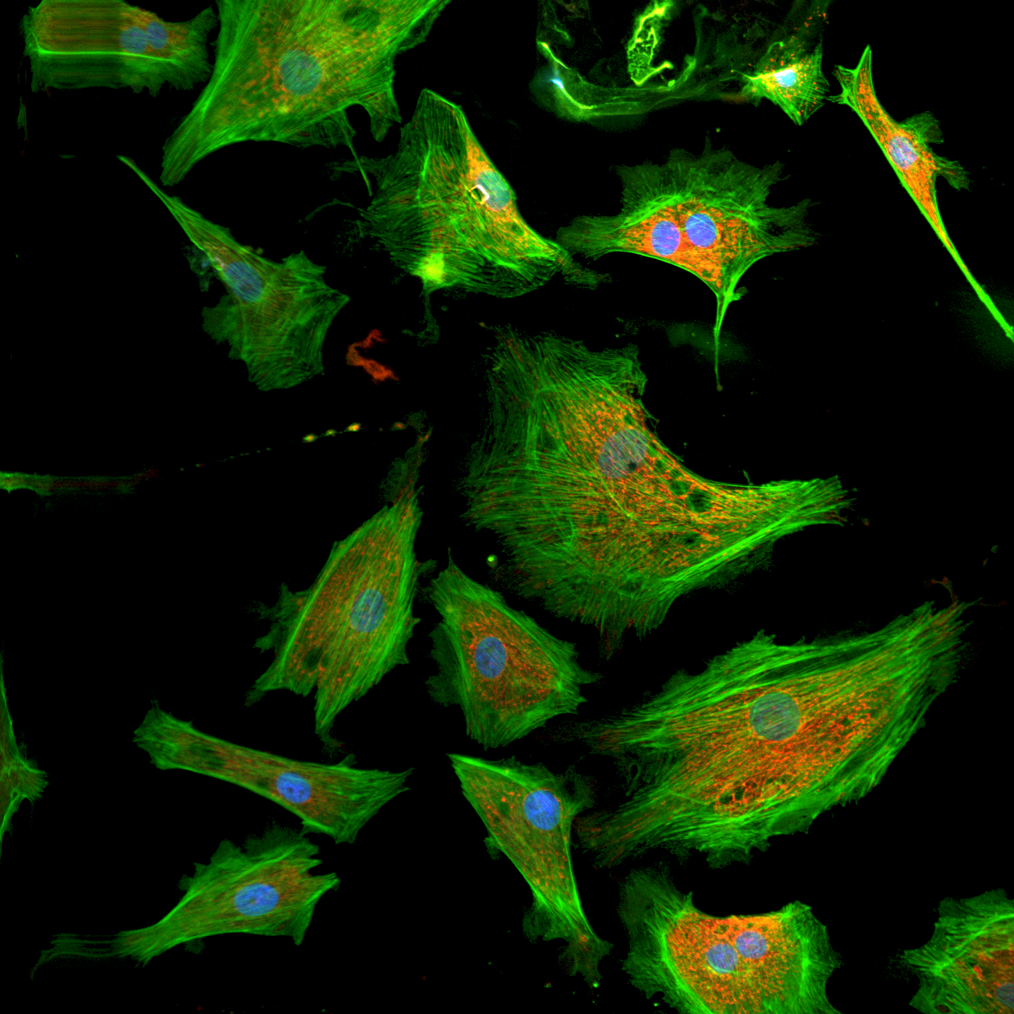
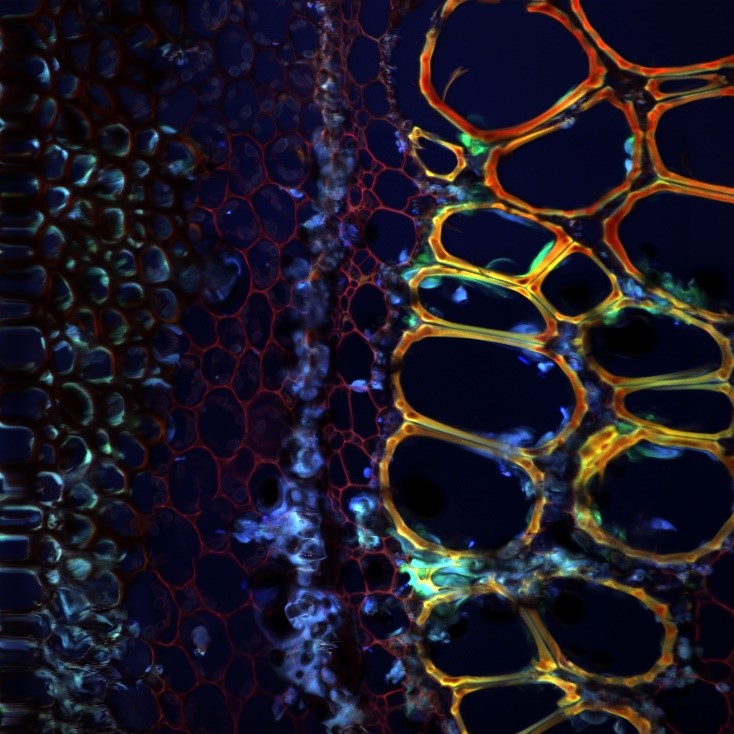
Sample Images
SEND MESSAGE